
The Laboratory of Cellular Pathology at Mater Dei Hospital Goes Digital
 11.07.2025
 5 minutes
 11.07.2025
5 minutes
11.07.2025
5 minutes
Formerly, medical students were trained to recognise different cell types in human tissues, enabling them to determine which organ was affected and to identify abnormalities, such as malignant cells indicative of cancer. This training often involved spending many hours examining cells under a microscope.
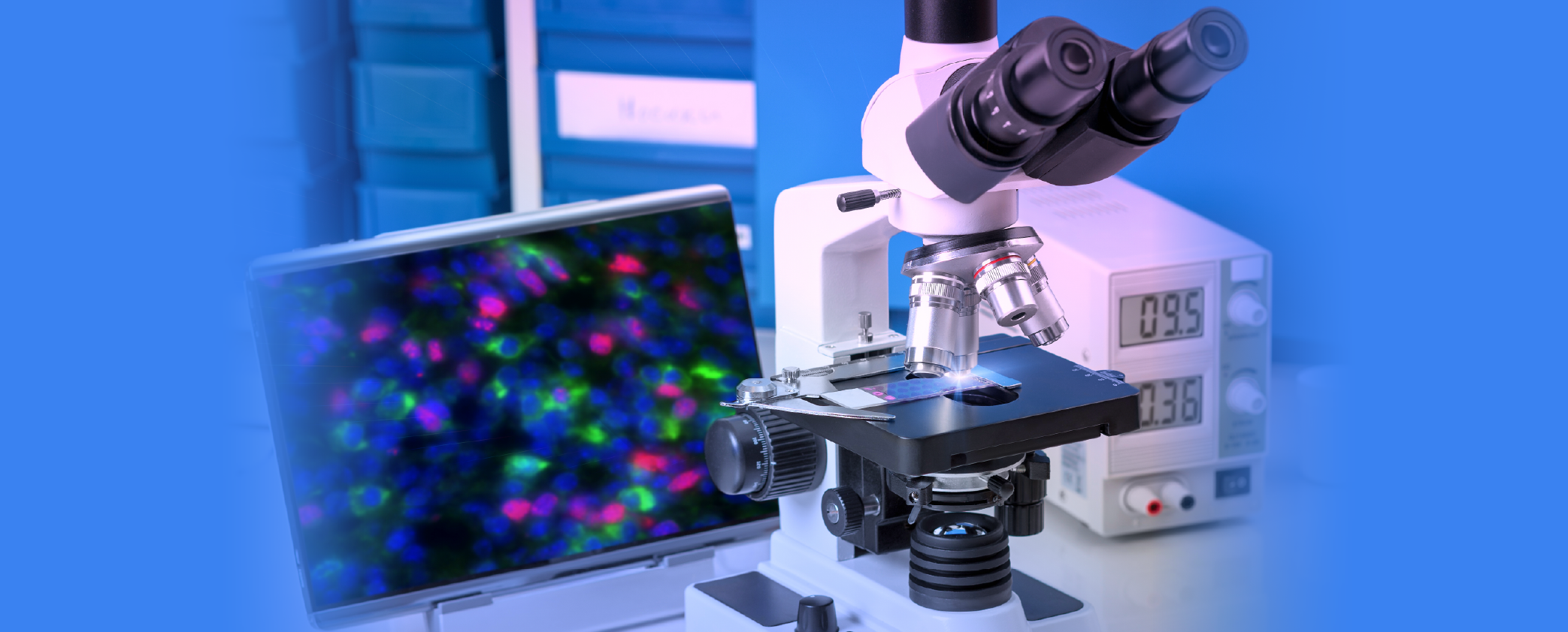
It was once unimaginable that a time would come when microscopes would be set aside, but that time has arrived. Today, tissues removed during surgery are processed in the Cellular Pathology Laboratory at Mater Dei Hospital. These samples are embedded in wax, sliced into ultra-thin sections, and placed on slides. From there, the pathologist - a specialist in cellular pathology - examines the cells' morphology and reports findings to the treating physician.
Thanks to this digital transformation, high-definition scanners are now used instead of traditional microscopes. These scanners generate detailed images of tissue samples, which are analyzed on 4K monitors. The digital images are crystal clear and can be zoomed, annotated, or compared using computer software.
If a pathologist needs to compare current findings with samples from years ago, there is no need to retrieve old physical slides or tissue blocks. All images are securely stored on the department’s digital server. As a result, reports are produced more rapidly and with improved accuracy. If a second opinion is needed from an expert abroad, digital files can be transferred instantly - eliminating the need to courier physical specimens. This process is now seamless and immediate.
Although this digital shift may appear straightforward, it presents certain challenges. High-resolution images, detailed enough to reveal individual cells, demand substantial digital storage. Each month, approximately seven terabytes of space are required to archive tissue images submitted by surgical colleagues. The scanners, monitors, computers, and servers used in this process are costly, with the total investment reaching €3 million. Fortunately, 80% of the funding came from European sources -specifically, the Recovery and Resilience Plan (C4-12), which supports the digitalization and modernization of the health system.
To further improve diagnostic precision and treatment planning, various immunohistochemical stains are applied. These offer critical insights into the most effective therapies for each individual case. This is the essence of personalised medicine - selecting targeted treatments that offer the highest chance of success, particularly in cancer care.
During a conference held as part of the Public Service Expo Village ’25, the successful completion of the digital pathology project was officially announced. The Minister for Health and Active Ageing, Jo Etienne Abela, explained how this new digital workflow is enabling faster, more personalised patient care, while also supporting a paperless and more sustainable healthcare environment through telepathology.
.png)
.png)


.png)